

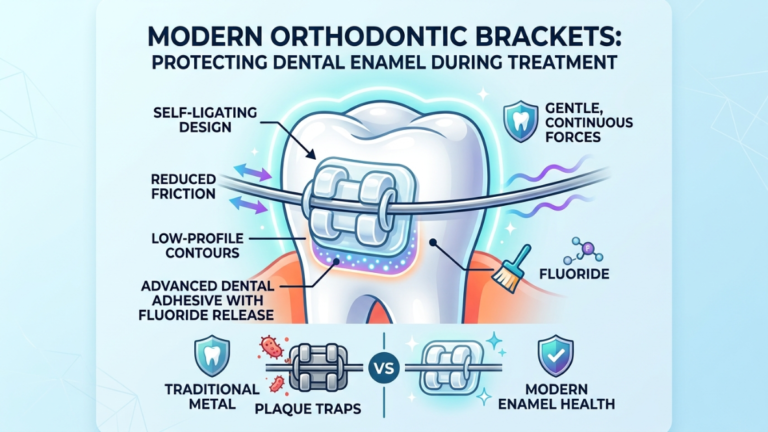
Ask any orthodontist who trained before the early 2000s about the quiet trade-off of straightening teeth, and they’ll usually bring up the same thing: faint white patches that sometimes appeared around brackets once the braces finally came off. Permanent reminders etched into the enamel by months of trapped plaque. That risk hasn’t disappeared, but it has shrunk considerably. Bracket systems, bonding chemistry, and even the technique used to remove brackets have all been re-engineered around one shared goal, move teeth into alignment without leaving a mark on the hardest tissue in the human body.
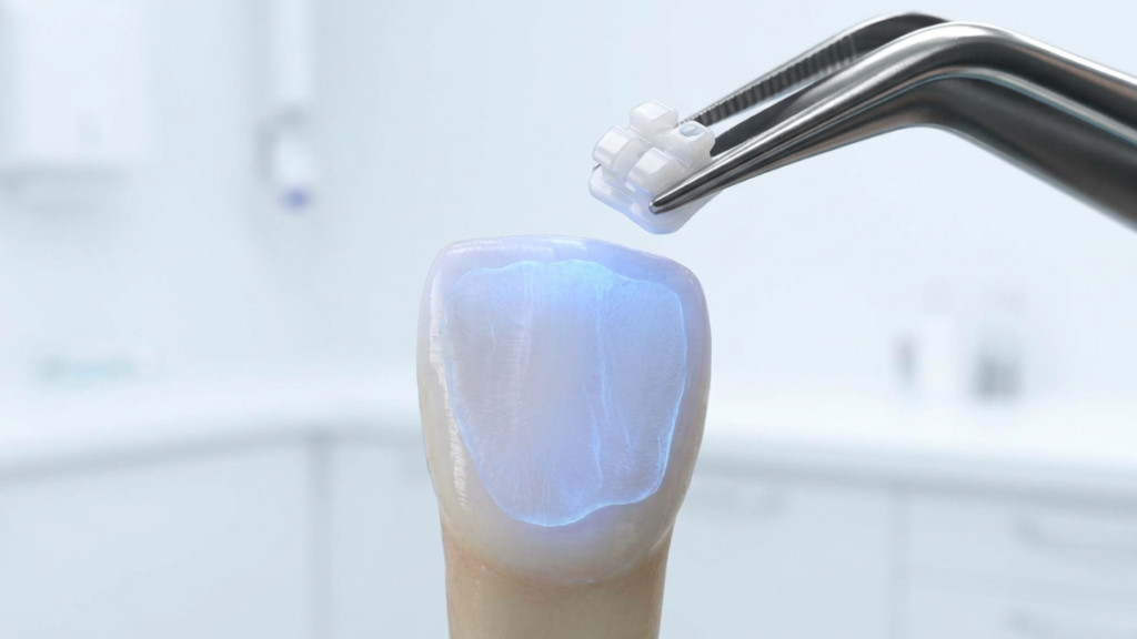
This guide breaks down exactly how that protection works, from the adhesive hiding under the bracket base to the method used to peel it off months later, and where genuine trade-offs still exist that rarely come up during a consultation.
Key Takeaways
- Enamel demineralization: white spot lesions is the most common enamel risk during orthodontic treatment, and it’s driven by plaque trapped against bracket surfaces, not by the bracket hardware itself.
- Fluoride-releasing adhesives, resin-modified glass ionomer cements, and bioactive glass fillers actively fight demineralization by releasing protective ions into surrounding enamel for the full duration of treatment.
- Self-ligating brackets reduce plaque-retentive surfaces by eliminating elastic ties, which lowers hygiene-related enamel risk though they don’t remove the need for diligent brushing.
- Ceramic brackets look gentler but are harder than natural tooth enamel, meaning they can wear down opposing teeth if a case isn’t planned carefully.
- Debonding, not just the months of active wear, is a genuine risk point for enamel; laser-assisted and low-force techniques now exist specifically to limit enamel loss when brackets and adhesive come off.
What Actually Happens to Enamel During Orthodontic Treatment?
Braces themselves don’t damage enamel directly, the real risk comes from acid-producing plaque bacteria that colonize rough surfaces around brackets. When sugars and starches feed that plaque, it releases acid that pulls calcium and phosphate out of enamel, a process called demineralization that can appear as chalky white spots within weeks.
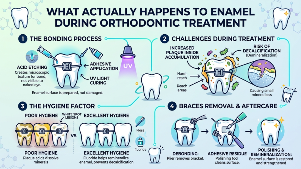
Enamel is built almost entirely from hydroxyapatite, a crystalline calcium-phosphate mineral that gives it exceptional hardness but also makes it acellular, it has no living cells and cannot heal itself the way skin or bone does. Whatever mineral enamel loses has to be replaced through an external chemical process called remineralization, where calcium and phosphate ions from saliva, or from a topical product, redeposit into the weakened crystal structure. There’s no biological repair crew doing this work; it’s pure chemistry, which is part of why prevention matters so much more here than in almost any other tissue in the body.
Fixed appliances change the geography of the tooth surface. Bracket bases, ligature grooves, and the margins where adhesive meets enamel all create tiny ledges and undercuts that a toothbrush bristle struggles to reach. Bacteria, particularly Streptococcus mutans, colonize those protected zones and build biofilm that a normal swipe of brushing won’t disturb. When that biofilm metabolizes dietary sugars, it drops the local pH low enough that hydroxyapatite starts dissolving. Do that repeatedly, several times a day, and the enamel around a bracket can visibly chalk within a matter of weeks, long before most patients would expect any change to be showing.
Why This Matters More With Braces Than Without
A patient with no fixed appliances has a much easier time keeping every tooth surface accessible to a toothbrush. Add fifteen to twenty brackets, an archwire, and possibly elastic ligatures, and a large share of enamel surface area sits behind hardware for twelve to twenty-four months at a stretch. That extended exposure window is precisely why bracket and adhesive design has become such a focused area of dental materials research over the past two decades, manufacturers were solving for the fact that hygiene alone, however diligent, wasn’t fully closing the gap.
How Do Modern Bonding Adhesives Actively Protect Enamel?
Many contemporary orthodontic adhesives are engineered to release fluoride, calcium, or phosphate ions directly into the surrounding enamel, creating a protective reservoir for the entire time brackets stay on,unlike older bonding resins, which were chemically inert once cured and offered enamel no ongoing defense.
This is arguably where the quietest revolution in bracket technology has happened. A conventional composite resin, once light-cured, is essentially plastic glued to a mineral surface, durable, but chemically passive. It holds the bracket in place and does nothing else. Ion-releasing adhesives change that equation by turning the bonding material itself into a slow-release protective agent for as long as it stays on the tooth.
Fluoride-Releasing Composites and Glass Ionomer Cements
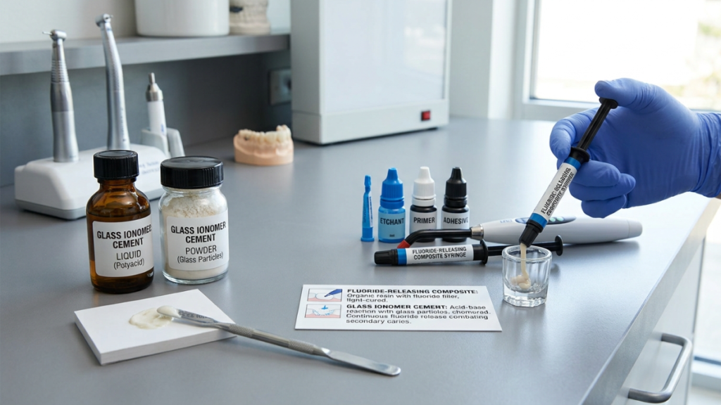
Resin-modified glass ionomer cements (RMGICs) and fluoride-releasing composite adhesives incorporate fluoride compounds that leach out gradually once bonded to the tooth. That fluoride gets taken up by the enamel immediately adjacent to the bracket base , precisely the zone most exposed to plaque acid , and helps convert some of the vulnerable hydroxyapatite into fluorapatite, a more acid-resistant mineral form. Laboratory research comparing bonded enamel exposed to a cariogenic environment consistently shows measurably less mineral loss around fluoride-releasing adhesives than around standard composite. Many of these materials can also be “recharged,” meaning their fluoride reservoir gets replenished every time the patient brushes with fluoride toothpaste or uses a fluoride rinse, which extends the protective effect across the full length of treatment instead of front-loading it in the first few weeks.
Bioactive Glass and Ion-Exchange Fillers
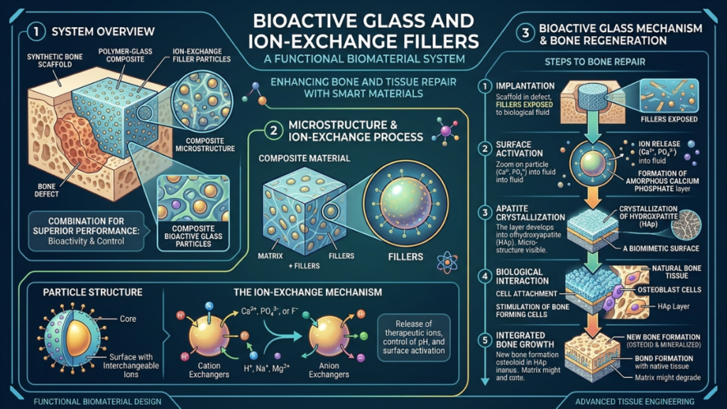
A newer category of adhesive filler uses bioactive glass, a calcium-sodium-phosphosilicate material originally developed for bone regeneration, blended into the bonding resin. When exposed to saliva, bioactive glass releases calcium, phosphate, and sodium ions that can precipitate into an apatite-like layer directly on the enamel surface, essentially building a mineral-rich buffer zone around the bracket. Because this filler acts as a reinforcing particle rather than a reactive resin component, it doesn’t meaningfully compromise shear bond strength, which has made it attractive to orthodontists who wanted ion release without sacrificing retention.
What’s on the Horizon: Antibacterial Coatings
The newest wave of research skips mineral chemistry and goes after the bacteria directly. Early-stage studies have coated brackets and adhesives with silver, zinc oxide, or titanium dioxide nanoparticles, all of which show antibacterial activity against the biofilm-forming species responsible for demineralization. Chitosan, a biodegradable compound derived from crustacean shells, has shown similar promise as a non-metallic antibacterial coating in laboratory testing. None of this is yet a mainstream standard of care, most of the supporting evidence still comes from in vitro testing rather than long-term clinical trials , but it signals where enamel-protective bracket technology is heading next: preventing the bacterial colony from establishing itself in the first place, rather than just resisting the acid it produces afterward.
Comparing Adhesive Systems
| Adhesive Type | Ion/Fluoride Release | Demineralization Protection | Shear Bond Strength | Best Suited For |
|---|---|---|---|---|
| Conventional composite resin | None | Baseline, no active protection | High, well-established | Low-risk patients with strong hygiene |
| Fluoride-releasing composite | Moderate, rechargeable | Meaningfully better than baseline | Comparable to conventional | General use, moderate caries risk |
| Resin-modified glass ionomer cement (RMGIC) | High, rechargeable | Strong protection near the bracket base | Slightly lower in some studies | Higher caries-risk patients |
| Bioactive glass-modified adhesive | Calcium, phosphate, sodium release | Builds a mineral-rich buffer zone | Maintained, minimal trade-off | Patients wanting ion release without bond compromise |
Does the Type of Bracket Affect Enamel Risk?
Yes. Bracket design changes how much plaque accumulates and how much force is needed to remove it later. Self-ligating brackets cut down on plaque-trapping elastic ties, while ceramic brackets, despite looking gentler, are harder than natural enamel and can wear down opposing teeth over time.
Self-Ligating vs. Conventional Ligation
Conventional brackets rely on small elastic ties, or less commonly thin stainless steel ligatures, to hold the archwire in its slot. Those elastic ties are porous, sit directly against the gumline, and are notorious for trapping food debris and plaque, they’re replaced at nearly every adjustment appointment partly for this reason. Self-ligating brackets use a built-in clip or sliding gate instead, so there’s no elastic surface for plaque to cling to and a cleaner overall profile for a toothbrush to reach. The clinical picture here is more nuanced than marketing materials sometimes suggest: self-ligating systems clearly produce less archwire friction and, in most comparisons, an easier hygiene profile, but recent narrative reviews conclude they don’t dramatically outperform conventional brackets across every treatment outcome. The realistic takeaway is that self-ligating design removes one specific plaque-retention risk factor, it isn’t a substitute for brushing.
Ceramic vs. Metal: A Hardness Mismatch Most Patients Don’t Hear About
Ceramic brackets, made from polycrystalline or monocrystalline alumina, are prized for blending in with natural tooth color. What rarely comes up in the consultation room is that alumina is harder than natural enamel. Laboratory wear testing has consistently found that ceramic brackets cause substantially more abrasive wear on opposing teeth than stainless steel brackets do when the two come into biting contact, a real concern for patients with a deep bite or an edge-to-edge tooth relationship, where a lower ceramic bracket might strike an upper front tooth repeatedly during normal chewing and speech. Orthodontists manage this by being selective about where ceramic brackets go, often reserving them for the upper arch only or avoiding them entirely in bite patterns where repeated contact is likely, rather than assuming the material is universally gentle just because it looks less industrial than metal.
Bracket Type Comparison
| Bracket Type | Plaque Retention Risk | Archwire Friction | Opposing-Tooth Wear Risk | Debonding Enamel Risk |
|---|---|---|---|---|
| Conventional metal (elastic tie) | Higher, elastic ties trap plaque | Higher | Low, metal-on-enamel contact is gentle | Low to moderate |
| Self-ligating metal | Lower, no elastic ties | Lower | Low | Low to moderate |
| Ceramic (self-ligating or conventional) | Moderate, depends on ligation type | Varies by system | Higher alumina is harder than enamel | Higher, more brittle, needs careful removal |
How Does the Bonding (Etching) Step Itself Affect Enamel?
Before bonding, enamel is conditioned with either traditional phosphoric acid etching or a milder self-etching primer. Self-etch primers remove less mineral and leave a shallower surface pattern, but several clinical trials show a modestly higher bond failure rate compared with conventional etching, a genuine trade-off, not a clear winner.
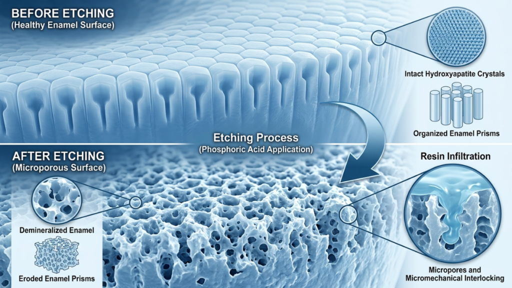
Traditional Phosphoric Acid Etching
The conventional protocol uses roughly 37% phosphoric acid applied for a short window, typically fifteen to thirty seconds, which dissolves a controlled amount of surface mineral and creates a honeycomb-like microscopic pattern that resin can mechanically lock into. It’s a well-understood, highly reliable technique with decades of clinical track record behind it, but it removes a measurable layer of surface enamel and, if handled inconsistently, can leave the subsurface structure slightly weakened right at the bracket-adhesive interface, exactly where forces concentrate later during debonding.
Self-Etch Primers: Enamel-Conserving, With a Catch
Self-etch primers combine the etchant and the priming agent into a single low-pH solution, cutting a step out of the bonding appointment and producing a noticeably shallower dissolution pattern under microscopic examination, meaning less enamel mineral is removed up front. That sounds like a straightforward win, and for enamel conservation alone, it largely is. The catch shows up in bond reliability: pooled data from multiple randomized trials shows a modest but real increase in bond failure risk with self-etch primers compared with conventional acid etching over a twelve-month follow-up period, meaning a slightly higher chance a bracket detaches and needs re-bonding. Orthodontists weigh this trade-off case by case, a patient with excellent compliance and low caries risk might be a great candidate for the gentler self-etch approach, while a higher-risk case might call for the extra retention security of conventional etching.
What Can Patients Actually Do to Protect Their Enamel During Treatment?
Consistent oral hygiene protects enamel more than any bracket or adhesive technology alone. Brush at the gumline and bracket edges twice daily with fluoride toothpaste, clean between brackets and wires daily, limit sugary or acidic snacking between meals, and keep every scheduled orthodontic check-up.
Daily Habits That Matter Most
No adhesive chemistry or bracket design fully compensates for inconsistent brushing,the technology narrows the margin for error, it doesn’t erase it. A soft-bristled or electric toothbrush angled at roughly 45 degrees toward the gumline, worked specifically along the top and bottom edges of every bracket, removes plaque that standard flat brushing tends to miss. Interdental brushes sized to slide under the archwire, or a water flosser on a gentler setting, reach the contact points between teeth that a bracket’s presence makes nearly impossible to floss conventionally. Fluoride toothpaste, used twice daily rather than rinsed away immediately afterward, gives enamel a steady low-dose supply of the exact mineral it needs to resist acid attack. On the diet side, the frequency of sugar and acid exposure matters more than the total amount consumed in a day, grazing on sugary drinks or snacks throughout the day keeps enamel in a near-constant acidic state, while the same sugar consumed in one sitting gives saliva a chance to buffer and recover in between.
Professional Remineralization Support
For patients already showing early signs of demineralization, orthodontic offices frequently apply topical fluoride varnish at routine visits to reinforce enamel between appointments. Some practices also recommend at-home casein phosphopeptide–amorphous calcium phosphate (CPP-ACP) paste, a milk-protein-derived remineralizing agent often recognized by product names like Tooth Mousse, which supplies bioavailable calcium and phosphate to weakened enamel. It’s worth being candid about the evidence here: while CPP-ACP has shown clear benefit over doing nothing, several systematic reviews have found its remineralizing effect isn’t consistently superior to fluoride alone, and combining the two appears to outperform either used in isolation. It’s a reasonable adjunct for higher-risk patients, not a replacement for fluoride or hygiene.
How Is Enamel Protected When Brackets Come Off?
Removing brackets and cleaning up leftover adhesive is one of the highest-risk moments for enamel in the entire treatment. Modern practices use controlled low-force pliers, fine-finishing burs or polishing discs instead of aggressive grinding, and increasingly laser-assisted techniques to loosen adhesive without touching the enamel surface at all.
Why Debonding Carries Real Risk
Debonding happens in two stages: prying the bracket itself off the tooth, then cleaning residual adhesive off the enamel surface underneath. Both stages can shave away surface enamel if performed aggressively. Studies quantifying this have found enamel loss during adhesive cleanup ranging from a few microns with careful, controlled technique up to considerably more when high-speed rotary instruments are used carelessly or held too long in one spot. Ceramic brackets raise the stakes further: their brittleness and higher fracture resistance mean more mechanical force is often needed to pop them free, which raises the risk of enamel chipping or delamination, a genuine reason some orthodontists steer patients toward metal in cases with an already-compromised bite.
Enamel-Conserving Removal Techniques
Clinicians grade cleanup quality using the Adhesive Remnant Index (ARI), a scale measuring how much resin is left on the tooth versus how much stayed on the bracket, which serves as a rough proxy for how conservatively the debond was performed. Fine-grit tungsten carbide burs and diamond polishing discs run at controlled, low speeds are now generally favored over aggressive grinding, since they remove residual composite without gouging into sound enamel underneath. For ceramic brackets specifically, laser-assisted debonding has emerged as a genuinely useful innovation: a controlled laser, commonly erbium or CO2-based, briefly softens the bond at the adhesive-enamel interface before mechanical removal, meaningfully reducing the force needed to pop the bracket free and, with it, the risk of enamel fracture.
Debonding Technique Comparison
| Removal Method | Enamel Loss Risk | Relative Speed | Best For |
|---|---|---|---|
| Low-speed tungsten carbide bur | Low to moderate, technique-dependent | Moderate | Most conventional adhesive cleanup |
| High-speed rotary bur, used carelessly | Higher | Fast | Not generally recommended for finishing |
| Diamond polishing discs | Low | Slower | Final finishing pass after bulk removal |
| Mechanical debonding pliers (ceramic) | Higher, brittle fracture risk | Fast | Standard ceramic bracket removal |
| Laser-assisted debonding | Low, softens bond before removal | Slower, more steps | Ceramic brackets, enamel-sensitive cases |
What Are the Biggest Misconceptions About Braces and Enamel Damage?
The most common myth is that ceramic or “natural-looking” brackets are automatically gentler on enamel. In reality, ceramic’s hardness can wear opposing teeth, self-etch bonding trades enamel conservation for a higher failure rate, and white spot lesions don’t fade away on their own, they need active remineralization.
“Ceramic is the safer choice.” As covered above, ceramic’s hardness relative to enamel is a genuine trade-off, not an upgrade in every respect. It’s cosmetically preferable for many patients, but “gentler on your teeth” isn’t automatically true.
“White spots will fade once the braces are off.” They generally won’t, not without help. A white spot lesion is a real loss of subsurface mineral, not a stain or surface discoloration that time erases on its own. Left alone, many lesions stay visible indefinitely and, in worse cases, progress toward actual cavitation. Early lesions respond well to fluoride and remineralizing pastes; more established ones sometimes need cosmetic intervention like resin infiltration or microabrasion afterward.
“Self-ligating brackets mean I don’t have to worry about cavities.” They lower one specific risk factor, plaque-trapping ties, but they don’t neutralize the need for hygiene. Plaque still accumulates around any bracket base, self-ligating or not.
“Braces mechanically wear down your teeth.” For the vast majority of patients, the archwire and bracket hardware itself doesn’t erode enamel through simple friction. The two documented mechanical exceptions are opposing-arch wear from unusually hard bracket materials like ceramic, and force applied during debonding, both real, both manageable, and both very different from the plaque-driven demineralization that accounts for most enamel changes during treatment.
“Remineralizing paste is basically a cure.” CPP-ACP and similar products are a genuinely useful adjunct, especially for patients at elevated risk, but the research doesn’t support treating them as a stand-alone fix that outperforms fluoride and hygiene. They work best layered on top of the fundamentals, not instead of them.
When Should You Be Concerned About Enamel Changes During Treatment?
Contact your orthodontist if you notice chalky white patches, rough spots, or new sensitivity near a bracket. These are early signs of demineralization, and they respond far better to fluoride varnish or remineralizing treatment when caught early, well before a lesion has any chance of becoming a cavity.
Mild, transient sensitivity after an archwire adjustment is normal and typically settles within a day or two, that’s tooth movement, not enamel damage. What’s worth flagging to your orthodontist is anything that looks like a persistent chalky or opaque patch near a bracket base, a rough texture you can feel with your tongue or fingernail, or sensitivity that’s new and doesn’t fade. Demineralization exists on a spectrum, and the earlier it’s caught, the more reversible it tends to be, a lesion identified in its first few weeks responds to fluoride varnish and improved hygiene far more predictably than one that’s had six months to deepen. Most orthodontic practices check for exactly this at every routine visit, but patients are in the mirror every day; catching something between appointments and mentioning it early is genuinely useful.
Frequently Asked Questions
Do braces permanently damage tooth enamel?
Not inherently. The hardware itself isn’t the source of damage for most patients, plaque accumulation around brackets is. With consistent hygiene, adequate fluoride exposure, and modern ion-releasing adhesives, most patients complete treatment with no lasting enamel change. Risk rises mainly with poor hygiene, prolonged treatment, or an aggressive debonding technique.
How quickly can white spot lesions form after getting braces?
Faster than most people expect. Visible chalky demineralization can begin appearing within about a month of bonding if plaque is allowed to build up around brackets and isn’t disturbed regularly. This is exactly why hygiene instruction typically starts on day one of treatment, not later.
Are self-ligating braces genuinely better for enamel health than traditional metal braces?
They offer a real but modest advantage. Removing elastic ties eliminates one plaque-retentive surface and generally makes brushing easier, which some research links to lower rates of decalcification-related complications. They aren’t dramatically superior across every outcome, though, and they still require the same hygiene commitment as any fixed appliance.
Can white spot lesions be reversed once braces come off?
Early-stage lesions often improve significantly with fluoride varnish, CPP-ACP paste, and time, since the mineral loss is still shallow enough for ions to redeposit. More advanced or long-standing lesions may not fully resolve on their own and sometimes benefit from cosmetic treatments like resin infiltration or microabrasion to blend the appearance back into the surrounding enamel.
Is some enamel loss during debonding unavoidable?
Some microscopic surface change is common even with careful technique, but it’s typically limited to the outermost polishing-level layer rather than anything structurally significant. The technique and instruments used matter far more than whether any change happens at all, controlled, low-force methods keep enamel loss to a level that’s clinically insignificant for nearly all patients.
Do ceramic braces really wear down other teeth?
Yes, in specific bite situations. Ceramic bracket material is harder than natural enamel, so a lower ceramic bracket that repeatedly contacts an upper tooth during chewing or speech can gradually wear that opposing enamel. Orthodontists screen for this during treatment planning and often avoid ceramic in bite patterns where this kind of contact is likely.
Visit this if you want know What is a Cavity Between Teeth?
