

What is an Interproximal Cavity?
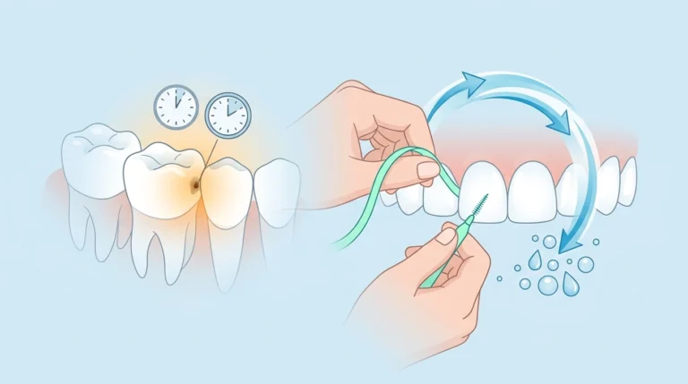
An Cavity Between Teeth ( interproximal cavity ) is a type of tooth decay that develops in the tight spaces between two adjacent teeth. It is primarily caused by plaque accumulation that cannot be reached by standard toothbrush bristles, leading to localized acid demineralization of the tooth enamel.
When you eat, food particles and sugars combine with oral bacteria—specifically Streptococcus mutans—to form a sticky biofilm known as plaque. Because the contact points between neighboring teeth are incredibly tight, this plaque remains trapped. Over time, the bacteria secrete acid byproducts that strip away the vital minerals (calcium and phosphate) from your tooth’s enamel shell, eventually forming a microscopic hole that expands inward toward the softer dentin layer.
Why Do Cavities Form Between the Teeth?
The Role of Interdental Plaque and Acid Byproducts
Standard toothbrushing only cleans the facial (front), lingual (back), and occlusal (chewing) surfaces of your teeth. This leaves up to 40% of your tooth surfaces uncleaned if you skip interdental hygiene. The stagnant plaque trapped in these tight contact zones continuously bathes the enamel in acid. This localized drop in pH quickly breaks down the hydroxyapatite crystal structure of your enamel, causing permanent structural failure.
Anatomical Risk Factors: Crowding and Tight Contact Points
Every individual possesses a unique oral anatomy that heavily influences their decay risk.
- Dental Crowding: Malaligned, overlapped, or severely crowded teeth create highly irregular spaces that trap debris and resist standard flossing paths.
- Broad Contact Areas: Some teeth meet at wide, flat surfaces rather than small points, expanding the surface area where plaque can securely anchor itself.
- Gingival Recession: As gum tissue recedes, it exposes the lower cementoenamel junction (CEJ). The root surface here is covered by cementum, which is significantly softer and less mineralized than enamel, making it twice as vulnerable to rapid interproximal decay.
Signs and Symptoms of a Hidden Cavity Between Teeth
Early Warning Signs (Asymptomatic Stage)
During the initial stages of an interproximal cavity, there are typically zero physical symptoms. Because the decay initiates between the teeth, it remains invisible to the naked eye during a standard mirror inspection. At this stage, only localized, faint white spots or minor structural shadows may be visible to a trained dentist.
Advanced Symptoms (Dentin and Pulp Involvement)
Once the acidic breakdown penetrates the protective enamel layer and reaches the underlying dentin, a patient will experience pronounced warning signs:
- Temperature Sensitivity: Sudden, lingering twinges of pain when consuming iced drinks, hot coffee, or highly sugary foods.
- Localized Pressure Discomfort: A sharp or dull ache localized to a specific quadrant when biting down or chewing hard foods.
- Chronic Halitosis: Persistent bad breath or a recurring foul taste caused by anaerobic bacteria processing trapped food debris within the interdental gap.
- Visual Shadows: A dark, grayish, or translucent shadow visible beneath the enamel surface of the chewing ridge adjacent to the interdental space.
The Floss Snag Test: A Warning Sign You Can Feel
One of the most reliable home indicators of a deteriorating interdental surface is changes in your daily flossing experience. If your dental floss consistently snags, frays, shreds, or gets physically stuck in a specific gap, it often signals that the enamel surface has broken open, creating a sharp, jagged shelf caused by a developing cavity cavity.
The “Kissing Cavity”: How Decay Spreads to Adjacent Teeth
A “kissing cavity” is a clinical term for two distinct interproximal cavities that form simultaneously on the touching surfaces of two adjacent teeth.
Because teeth rest in direct structural contact with one another, an active bacterial colony established on one tooth wall directly exposes the neighboring tooth wall to the exact same acidic microenvironment. The continuous accumulation of destructive plaque within that singular, shared boundary space means that an untreated interproximal cavity will almost always migrate laterally, causing a mirror-image cavity to form on the healthy adjacent tooth.
How Do Dentists Detect Cavities in Between Teeth?
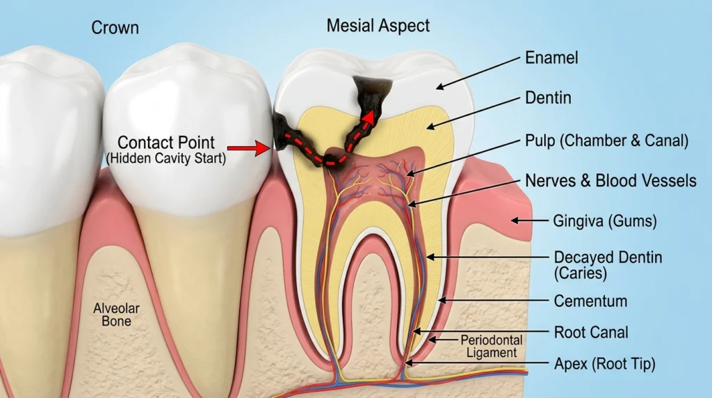
Dentists primarily detect cavities between teeth using bitewing X-rays, which reveal decay hidden beneath the enamel surfaces. Visual examinations and advanced near-infrared transillumination (NIRI) technologies are also utilized to catch early-stage lesions.
[Healthy Enamel] ---> [Incipient Decay] ---> [Dentin Intrusion] ---> [Pulpitis]
(No Action Required)  (Remineralization)    (Class II Filling)     (Root Canal)
Bitewing X-rays vs. Visual Dental Exams
A visual exam using a standard dental mirror and explorer tool can only detect an interproximal cavity if the decay has progressed so far that the structural ceiling of the tooth has completely collapsed. Bitewing radiographs (X-rays) are the absolute gold standard for early discovery. On a radiographic image, healthy enamel appears bright white, while active interproximal decay appears as a dark, triangular shadow penetrating the tooth wall.
Advanced Transillumination Technology (NIRI)
Modern dental practices use specialized Near-Infrared Transillumination (NIRI) diagnostic tools. This technique shines a specific wavelength of safe light directly through the structural framework of the tooth. Healthy enamel structures allow the light to pass cleanly, while areas of active mineral loss or early-stage interproximal lesions scatter the light, casting a clear diagnostic silhouette on a digital sensor without exposing the patient to radiation.
Treatment Options for Interproximal Tooth Decay
Incipient Decay: Remineralization and Silver Diamine Fluoride (SDF)
If an interproximal cavity is identified early—meaning it is strictly confined to the outer half of the enamel and has not breached the dentin barrier—it is classified as an incipient lesion.
At this stage, drilling is completely unnecessary. The tooth can be systematically saved through non-invasive remineralization therapies:
- Prescription-Strength Fluoride: Clinically applied varnishes high in sodium fluoride help pull calcium and phosphate minerals back into the porous enamel matrix.
- Silver Diamine Fluoride (SDF): A specialized liquid antimicrobial painted between the teeth that instantly kills cariogenic bacteria and halts further structural progression.
Moderate Decay: Class II Composite Fillings and Matrix Bands
Once the decay crosses the enamel-dentin junction, mechanical intervention is mandatory. Because the cavity resides on the side wall of the tooth, a dentist must access it by removing a small portion of the upper chewing surface. This procedure is called a Class II restoration.
To rebuild the missing side wall and ensure food cannot trap there later, the dentist utilizes a specialized ultra-thin metal or plastic strip known as a matrix band. This band is wrapped securely around the tooth during the application of tooth-colored composite resin, acting as a temporary mold to re-establish a perfectly smooth, tight contact point against the neighboring tooth surface.
Severe Decay: Inlays, Onlays, and Root Canal Therapy
If an interproximal cavity is left ignored, it will quickly migrate down into the deeper structures of the tooth:
| Treatment | Clinical Indication | Diagnostic Requirement |
| Inlay / Onlay | Structural decay is too extensive for a standard filling but does not require a full crown. | Custon laboratory-fabricated porcelain or gold insert. |
| Root Canal Therapy | The decay breaches the internal pulp chamber, causing irreversible infection or tissue death. | Complete removal of infected nerve tissues followed by biological sealing. |
| Dental Crown | The structural walls are severely compromised following a root canal or major structural fracture. | Full-coverage protective porcelain cap to restore functional bite forces. |
How Long Do You Have Before an In-Between Cavity Worsens?
The exact timeline for an interproximal cavity to progress through a tooth depends heavily on diet, salivary flow, and systemic hygiene habits.
The Clinical Timeline Rule: On average, it takes approximately 6 to 18 months for a cavity to cross the dense outer enamel layer. However, once the decay reaches the softer, highly porous dentin layer, the rate of destruction increases up to twice as fast.
At this juncture, a simple cavity can quickly transform into a painful nerve infection within a matter of months.
Preventive Interdental Home Care Strategies
To protect your smile from developing recurring interproximal decay, implement a dedicated daily interdental hygiene regimen:
- Floss with Proper Technique: Do not simply snap the floss down between your teeth. Curve the floss into a distinct “C-Shape” around the neck of each tooth, sliding it gently beneath the gumline to manually scrape away the sticky biofilm.
- Incorporate Interdental Brushes: If you have wider gaps, localized gum recession, or orthodontic appliances, use small, specialized interdental brushes (proxabrush). Clinical trials show these brushes are highly efficient at removing stubborn interproximal plaque.
- Use a Target Hydro-Flosser: Utilize a high-quality water flosser to power away loose food debris and flush out active bacteria hidden deep inside the interdental pockets.
- Rinse with Therapeutic Mouthwash: Incorporate an alcohol-free, anticavity mouthwash formulated with sodium fluoride or essential oils to neutralize residual acids and remineralize hard-to-reach contact areas.
Frequently Asked Questions About Interproximal Cavities
Can a cavity between teeth heal on its own?
A cavity between teeth can only heal on its own if it is caught in the early “incipient” stage, where decay is confined strictly to the outer enamel. At this stage, it can be remineralized using prescription fluoride treatments and meticulous interdental hygiene. Once decay breaches the softer dentin layer, it cannot heal naturally and requires a professional filling.
Why does my floss always rip or tear in the same spot?
When dental floss consistently tears or frays in a specific spot, it is usually caused by a sharp ledge or structural hole in the tooth enamel created by an underlying cavity, or a failing, jagged edge of an old dental filling.
Do fillings between teeth feel natural?
Yes. By using advanced matrix bands and anatomical wedges, dentists can sculpt modern composite resin materials to mirror the exact natural curvature of your original tooth structure, preserving a comfortable, unnoticeable bite.
